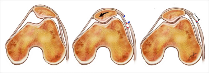
Figure 6.
Illustrations showing the axial views of the patellofemoral joint and the effect of lateral retinacular lengthening. On the left, the original patellofemoral joint shows lateral tilt of the patella. On the center, the superficial (blue) and deep (red) layers of the lateral retinaculum have been incised with the correction of the lateral tilt. On the right, the superficial and deep layers are sutured lengthening the lateral retinaculum.

