Figure 3.
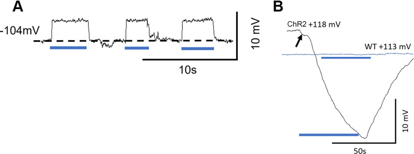
Light activation of ChR2 depolarizes ChR2 mouse DC membrane potentials in vivo. A, Examples of intracellular recordings from presumed DCs showing successive membrane depolarizations to OoC illumination. B, Prolonged OoC illumination and activation of ChR2 channels expressed in DCs and OPCs cause a large, slow reduction in the EP recorded from scala media during OoC illumination. ChR2 trace arrow indicates onset step. WT littermate (blue trace): nonresponsive to OoC illumination. A, B, Blue bars represent periods of illumination with a laser beam (200 µm diameter, 0.25 mW mm−2, 470 nm).

