Figure 1.

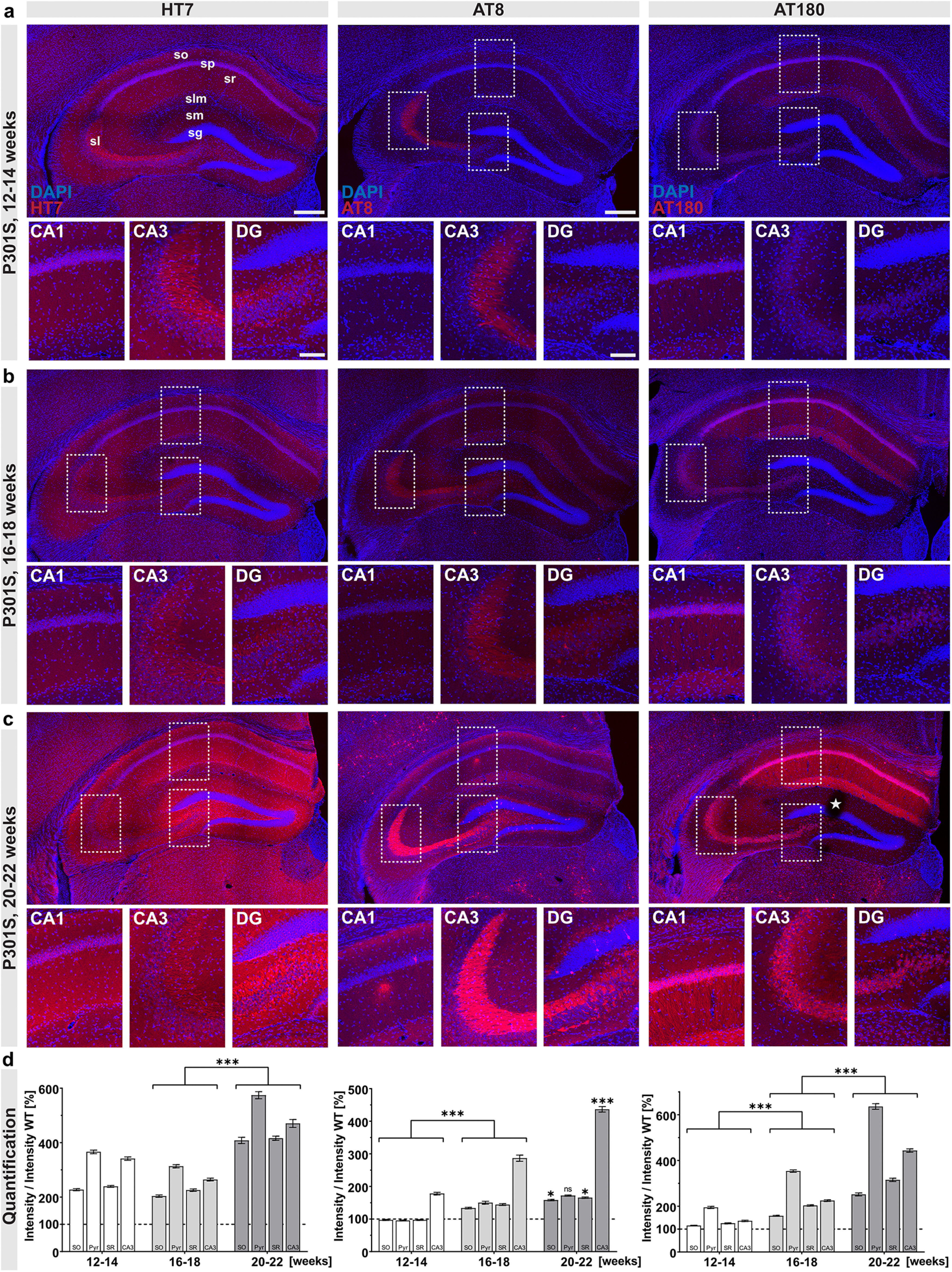
Progressive Tau pathology in P301S mice. Overview and zoom-in (CA1, CA3, DG) of the hippocampus of P301S mice. a, At 12-14 weeks of age, immunoreactivity with the HT7 antibody, detecting total hTau, was present in all hippocampal subfields. AT8 immunoreactivity was mainly observed in stratum lucidum (sl), while AT180 immunoreactivity was present at low levels in the stratum pyramidale (sp) of the CA1-CA3 region and to a lesser extent in the stratum oriens (so), stratum radiatum (sr), and lacunosum moleculare (slm). b, At the age of 16–18 weeks, all hippocampal subfields show HT7 immunoreactivity. A weak AT8 immunoreactivity was detected in all layers of the CA1 region as well as in the DG, where AT8+ cells were present in the hilus. Immunoreactivity for AT180+ Tau increased in all layers of the CA1-CA3 region compared with 12–14 weeks. c, At the latest time point analyzed (20–22 weeks), HT7 immunoreactivity massively increased in all hippocampal subfields. AT8+ cells were detected throughout the CA1 region, with a massive accumulation of AT8 immunoreactivity in the mossy fibers of sl. In the DG, AT8+ cells were present in the hilus and the stratum granulosum (sg), showing a somatodendritic staining pattern. Somata and dendrites of CA1 pyramidal neurons show strong immunoreactivity for AT180, also somata of CA3 neurons show AT180 staining. d, Quantification of HT7, AT8, and AT180 immunoreactivity in hippocampal regions over time. Staining intensity in the four subregions was quantified relative to the respective earlier time points. AT8 (center) and AT180 (right) immunoreactivity significantly increased in all subregions from 12–14 to 16–18 weeks (AT8: 12–14 vs 16–18 weeks, SO, Pyr, SR, CA3, ***p < 0.0001; AT180: 12–14 vs 16–18 weeks, SO, Pyr, SR, CA3, ***p < 0.0001), while HT7 immunoreactivity was not altered (not significant.) From 16-18 to 20-22 weeks, all markers (HT7, AT8, AT180) showed a significant increase, except for AT8 immunoreactivity in the pyramidal cell layer (HT7: 16–18 vs 20–22 weeks, PO, Pyr, SR, CA3, ***p < 0.0001; AT8: 16–18 vs 20–22 weeks, SO, SR, *p < 0.05, CA3, ***p < 0.0001, Pyr not significant; AT180: 16–18 vs 20–22 weeks, SO, Pyr, SR, CA3, ***p < 0.0001). Background staining from WT slices was set to 100% (dashed line). Data are mean ± SEM. Intensities for each subregion were analyzed using either one-way ANOVA with Bonferroni's post hoc test, or using Kruskal–Wallis test with Dunn's test: *p < 0.05; ***p < 0.0001. All sections were imaged with constant laser intensity to visualize progressing Tau pathology. N = 3 animals per genotype and age, maximum intensity projections of representative confocal images, 40 µm coronal sections. Scale bars: hippocampal overview, 300 µm, zoom-in CA1, CA3, and DG, 100 µm. *Slicing artifact.
