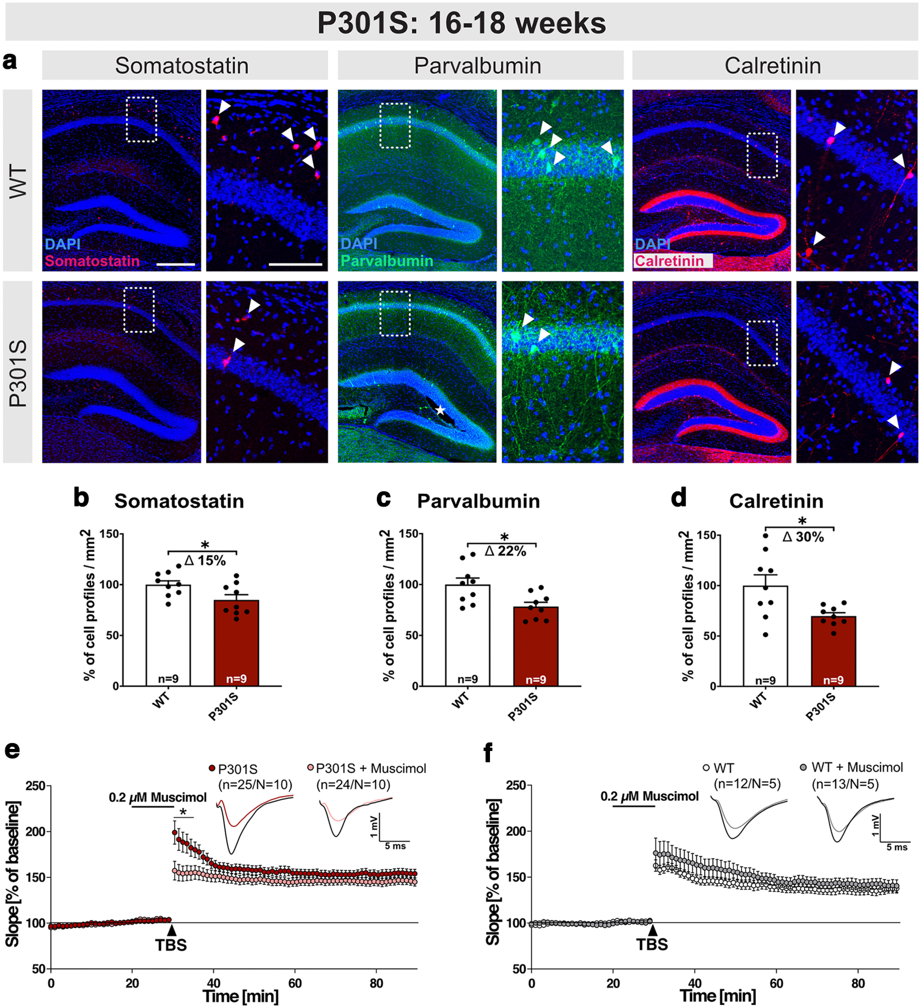
Figure 6.

P301S mice exhibit a loss of interneurons at 16-18 weeks. a, Representative coronal brain sections (40 µm) depicting the hippocampus of WT and P301S mice at 16-18 weeks of age. Boxed areas of CA1 are displayed at higher magnification on the right. SST+, PV+, and CR+ inhibitory interneurons were identified using specific antibodies. Arrowheads indicate exemplary cells for each marker. b–d, Hippocampi from P301S mice show a loss of SST+, PV+, and CR+ cell profiles/mm2 (SST: *p = 0.0301; PV: *p = 0.0118; CR: *p = 0.0156) compared with WT controls. e, f, Application of muscimol reduced LTP induction in P301S slices, while LTP in WT slices was not significantly different. Age of mice: 16-18 weeks Scale bars (a): overview, 300 µm; zoom-in, 100 µm; 10× mosaic confocal images, maximum intensity projections, slicing artifact marked with an asterisk. b–d, Data are mean ± SEM. n = number of sections from N = 3 mice/genotype, as indicated in the bar graphs. e, f, n = number of recorded slices; N = number of animals. Data were analyzed by Student's t test: *p < 0.05; **p < 0.01; ***p < 0.001.
