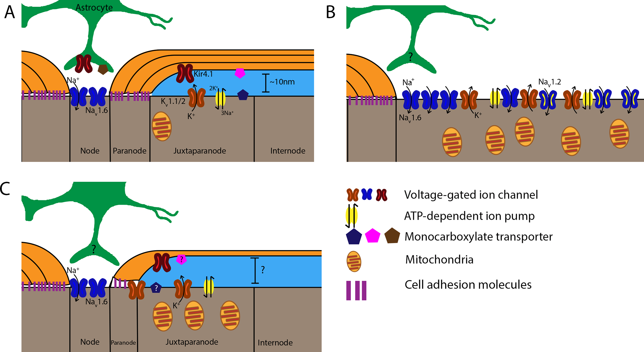
Figure 1: Nodal alterations following demyelination and remyelination.

Healthy axo-myelin units are displayed on the left of each panel for comparison. A: In healthy myelinated axons, voltage-gated sodium channels (Nav1.6, blue) are located at the node of Ranvier, while voltage-gated potassium channels (Kv1.1,2, orange), ATP-dependent Na+/K+ pumps (yellow), and mitochondria localize to the juxtaparanode. Inward-rectifying K+ channels (Kir4.1, red) are expressed by both oligodendrocytes and astrocytes, which extend perinodal processes that contribute to node function. Different monocarboxylate transporters (MCTs) are expressed by oligodendrocytes (MCT1, pink), axons (MCT2, blue), and astrocytes (MCT4, brown), indicating an important role for lactate metabolism. B: Loss of paranodal junctions (purple) due to demyelination results in Kv channel diffusion along the axolemma. Nav clusters also diffuse, and axons begin expressing Nav1.2 along the former internode. This increases the area across which ion gradients must be maintained, causing broad expression of Na+/K+ pumps and increasing mitochondrial density to meet axonal energy demands. Axonal MCT expression is reduced, potentially to compensate for high extracellular lactate levels. C: Following remyelination, ion channel and pump localization is restored, with some Kv channels persisting in the paranodal region. Mitochondrial density remains elevated, while the expression of MCTs during this period is unknown. Notably, the presence or absence of perinodal astrocyte processes during demyelination and remyelination has yet to be elucidated. Other morphological factors of remyelinated axons, such as the size of the periaxonal space, also remain unknown.
