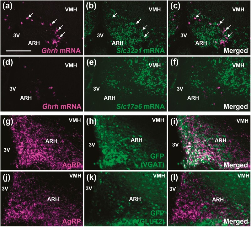
Figure 3.
Part of ARHGHRH neurons and practically all ARHAgRP neurons are GABAergic. A to C, Colocalization between Ghrh mRNA (magenta staining) and Slc32a1 mRNA (green staining) in the ARH (n = 4). The arrows indicate double-labeled neurons. D to F, No colocalization between Ghrh mRNA (magenta staining) and Slc17a6 mRNA (green staining) in the ARH (n = 4). G to I, ARHVGAT neurons (GFP staining in green representing VGAT-expressing cells) colocalize with AgRP peptide (magenta staining; n = 2). J to L, ARHVGLUT2 neurons (GFP staining in green representing VGLUT2-expressing cells) do not express AgRP (n = 2). Scale bar = 100 µm. 3V, third ventricle; AgRP, agouti-related protein; ARH, arcuate nucleus; GFP, green fluorescent protein; mRNA, messenger RNA; VGAT, vesicular inhibitory amino acid transporter; VMH, ventromedial nucleus.

