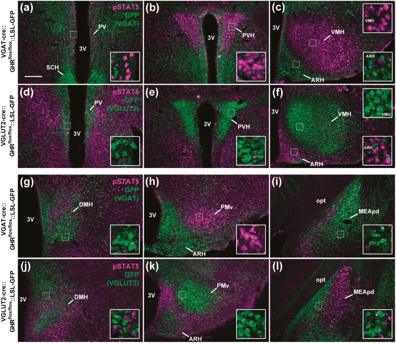
Figure 4.
GHR ablation in GABAergic and glutamatergic neurons. A to C and G to I, GABAergic neurons (VGAT-positive cells; GFP staining in green) are no longer responsive to GH in VGAT GHR KO mice, whereas GH-induced pSTAT5 (magenta nuclear staining) is still observed in non-GABAergic neurons (n = 3). D to F and J to L, Glutamatergic neurons (VGLUT2-positive cells) are no longer responsive to GH in VGLUT2 GHR KO mice, whereas GH-induced pSTAT5 is still observed in nonglutamatergic neurons (n = 3). Scale bar = 200 µm. Insets represent higher magnification images. 3V, third ventricle; ARH, arcuate nucleus; DMH, dorsomedial nucleus; GH, growth hormone; GHR, GH receptor; KO, knockout; MEApd, posterodorsal medial nucleus of the amygdala; opt, optic tract; PMv, ventral premammillary nucleus; PV, periventricular nucleus; PVH, paraventricular nucleus of the hypothalamus; SCH, suprachiasmatic nucleus; VGAT, vesicular inhibitory amino acid transporter; VMH, ventromedial nucleus.

