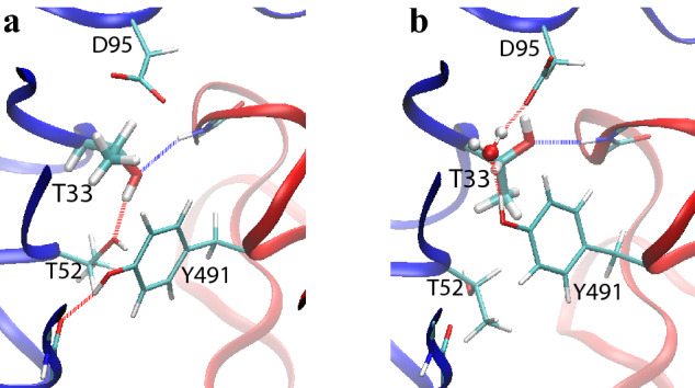
Figure 5.

Snapshots of two alternative side chain conformations for the residues around Thr33 in the binding interface between the m396 heavy chain (blue) and the SARS-CoV-1 RBD (red). H-bonds between the residues are shown. In panel (b), a water molecule is also involved in the H-bond network. A proper equilibration between these alternative conformations would require long simulation times, thus resulting in slow convergence of the FEP calculations. Images were rendered by VMD43 (version 1.9.3).
