(
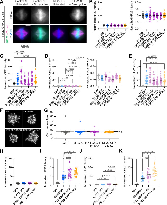
A) Immunofluorescence images of HeLa-Kyoto cells expressing KIF22-GFP under the control of a doxycycline inducible promoter. Images are maximum intensity projections in z of five frames at the center of the spindle. Fixed approximately 24 hr after siRNA transfection and treatment with doxycycline to induce expression. Scale bar 5 μm. KD: knockdown. (
B–E) Quantification of KIF22 fluorescence intensity in untreated HeLa-Kyoto cells transfected with control siRNA (
B), cells treated with doxycycline to induce expression and transfected with control siRNA (
C), untreated cells transfected with KIF22 siRNA (
D), and cells treated with doxycycline and transfected with KIF22 siRNA (
E) normalized to the mean intensity of uninduced, control knockdown cells (endogenous KIF22 expression level) for each cell line (
B). Data in (
B, D) are presented with the same y-axis scale as data in (
C, E) for comparison (left), and with independently scaled y-axes to show data variability (right). Twenty-seven GFP, 24 KIF22-GFP, 27 KIF22-GFP R149Q, 28 KIF22-GFP P148L, 25 KIF22-GFP P148S, 27 KIF22-GFP R149L, and 30 KIF22-GFP V475G untreated cells transfected with control siRNA (
B), 24 GFP, 24 KIF22-GFP, 31 KIF22-GFP R149Q, 30 KIF22-GFP P148L, 27 KIF22-GFP P148S, 30 KIF22-GFP R149L, and 33 KIF22-GFP V475G doxycycline-treated cells transfected with control siRNA (
C), 21 GFP, 31 KIF22-GFP, 27 KIF22-GFP R149Q, 32 KIF22-GFP P148L, 22 KIF22-GFP P148S, 22 KIF22-GFP R149L, and 25 KIF22-GFP V475G untreated cells transfected with KIF22 siRNA (
D), 26 GFP, 26 KIF22-GFP, 32 KIF22-GFP R149Q, 28 KIF22-GFP P148L, 28 KIF22-GFP P148S, 27 KIF22-GFP R149L, and 33 KIF22-GFP V475G doxycycline-treated cells transfected with KIF22 siRNA (
E) from three experiments. (
F) DAPI-stained metaphase chromosome spreads from uninduced RPE-1 cell lines with inducible expression of GFP, KIF22-GFP, KIF22-GFP R149Q, or KIF22-GFP V475G. Scale bar 10 μm. Images are representative of three experiments. (
G) Numbers of chromosome pairs counted in metaphase spreads prepared from RPE-1 stable cell lines. Dashed line indicates the expected chromosome number for diploid human cells (46). The mode for each cell line is 46. Fifty-five GFP, 58 KIF22-GFP, 53 KIF22-GFP R149Q, and 57 KIF22-GFP V475G spreads from three experiments. (
H–K) Quantification of KIF22 fluorescence intensity in untreated RPE-1 cells transfected with control siRNA (
H), cells treated with doxycycline to induce expression and transfected with control siRNA (
I), untreated cells transfected with KIF22 siRNA (
J), and cells treated with doxycycline and transfected with KIF22 siRNA (
K) normalized to the mean intensity of uninduced, control knockdown cells for each cell line (
H). Twenty-three GFP, 27 KIF22-GFP, 25 KIF22-GFP R149Q, and 27 KIF22-GFP V475G untreated cells transfected with control siRNA (
H), 24 GFP, 27 KIF22-GFP, 27 KIF22-GFP R149Q, and 28 KIF22-GFP V475G doxycycline-treated cells transfected with control siRNA (
I), 21 GFP, 24 KIF22-GFP, 24 KIF22-GFP R149Q, and 21 KIF22-GFP V475G untreated cells transfected with KIF22 siRNA (
J), 24 GFP, 29 KIF22-GFP, 26 KIF22-GFP R149Q, and 24 KIF22-GFP V475G doxycycline-treated cells transfected with KIF22 siRNA (
K) from three experiments. For (
B–E) and (
H–K), bars indicate means. P values from Brown-Forsythe and Welch ANOVA with Dunnett’s T3 multiple comparisons test. P values are greater than 0.05 for comparisons without a marked p value. See
Figure 2—figure supplement 1—source data 1.


