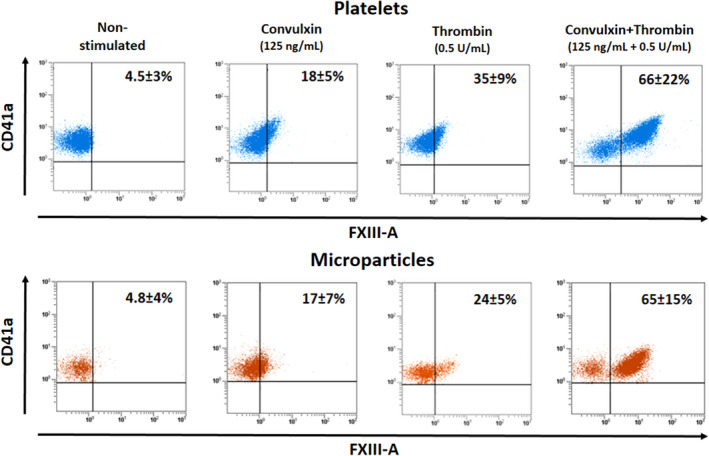
FIGURE 2.

Surface exposure of factor XIII A subunit (FXIII‐A) on platelets activated by receptor mediated mechanism and on the formed microparticles. In each case platelets were activated for 15 min. The detection of CD41a antigen was used for the identification of platelets and platelet‐derived microparticles. Microparticles were gated at 1 µm. The upper row demonstrates the results with activated platelets, while in the lower row results obtained with the formed microparticles are shown. The dot plots show the results of representative individual samples, while the numbers in the right upper corners represent the means ± standard deviations; each value was calculated from measurements on samples from five different donors. With the exception of thrombin activation there was no significant difference between FXIII‐A positivity of platelets and microparticles. In the case of thrombin induced activation, the difference of platelets versus microparticles was significant (P = .038)
