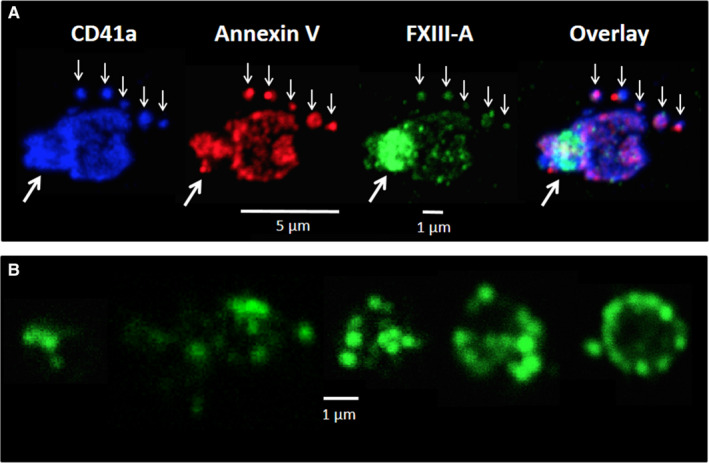
FIGURE 3.

Surface labeling of platelets activated by receptor mediated mechanism and of the formed microparticles using immunofluorescent markers. A, Platelet activated by convulxin+thrombin stained for CD41a (blue), annexin V binding (phosphatidylserine labeling, red) and factor XIII A subunit (FXIII‐A; green). The small white arrows point to microparticles, while the thick white arrows point to the cap‐like structure on an activated platelet. B, FXIII‐A positive clumped microparticles formed from platelets activated by convulxin+thrombin. The images are representative of experiments performed on platelets from three different donors
