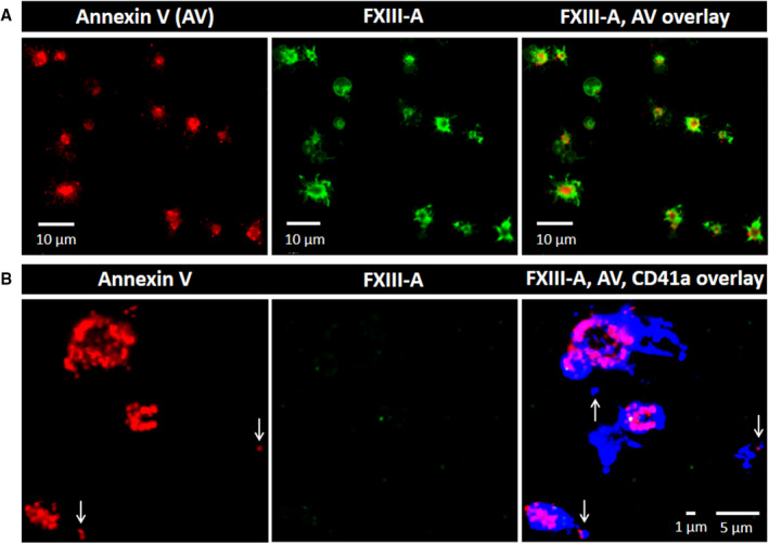
FIGURE 6.

Labeling of permeabilized and non‐permeabilized platelets activated by Ca2+‐ ionophore. Platelets were stimulated with 0.7 µM calcimycin for 15 min. A, Platelets were permeabilized and labeled for phosphatidylserine (annexin V binding; red) and factor XIII A subunit (FXIII‐A; green). B, Non‐permeabilized platelets were stained for annexin V binding, FXIII‐A and CD41a (blue). Vertical arrows indicate microparticles. Note the absence of FXIII‐A staining on non‐permeabilized platelets as opposed to permeabilized ones. The images are representative of experiments performed with platelets from three different donors
