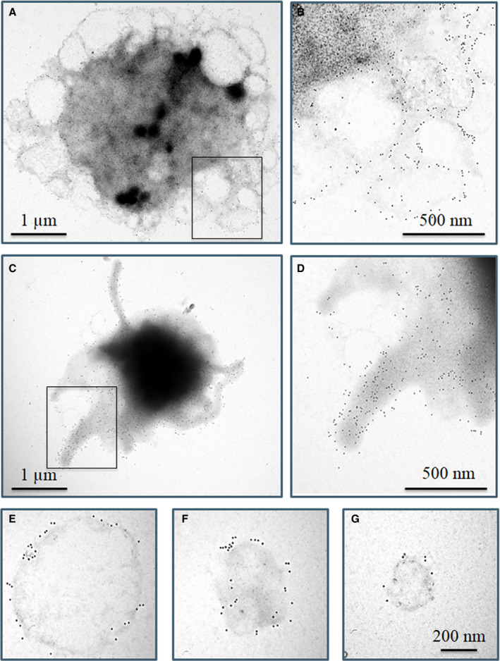
FIGURE 7.

Surface labeling of Ca2+‐ionophore activated platelets using immunogold technique. Platelets were stimulated with 0.7 µM calcimycin. A‐D, Two different cellular structures of calcimycin activated platelets were observed, both of which were labeled for CD41a (10 nm gold particles). A,C, Whole platelets, (B,D) higher magnification of selected areas from (A,C). The absence of 15 nm gold particles on either platelet morphology indicates the lack of labeling for factor XIII A subunit (FXIII‐A). E–G, Microparticles of various size intensively labeled for CD41a failed to label for FXIII‐A. The images are representative of experiments performed with platelets from three different donors
