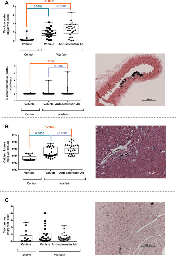
Fig. 5.

Anti‐sclerostin antibody treatment leads to increased development of vascular calcifications. Calcium content of (A, upper panel) the aorta, (B) kidney, and (C) heart of mice treated with anti‐sclerostin antibody or vehicle after exposure to warfarin‐containing or control diet, accompanied by representative images of von Kossa‐stained tissue sections of anti‐sclerostin antibody‐treated mice (counterstained with H&E). (A, lower panel) Quantification of the calcified area as measured on von Kossa–stained tissues sections. These calcifications are located in the medial layer of the aorta, following the elastic lamellae. (B) Areas of calcification in the kidney were confined to the vasculature. (C) Spontaneous calcifications developed in the myocardium of DBA/2 mice.
