FIGURE 3.

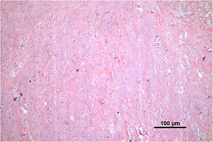
Necrosis of periprosthetic tissue. Paler stained, cell‐free tissue area, as compared to more cell‐rich areas as shown in Figure 1. H&E, 20×

Necrosis of periprosthetic tissue. Paler stained, cell‐free tissue area, as compared to more cell‐rich areas as shown in Figure 1. H&E, 20×