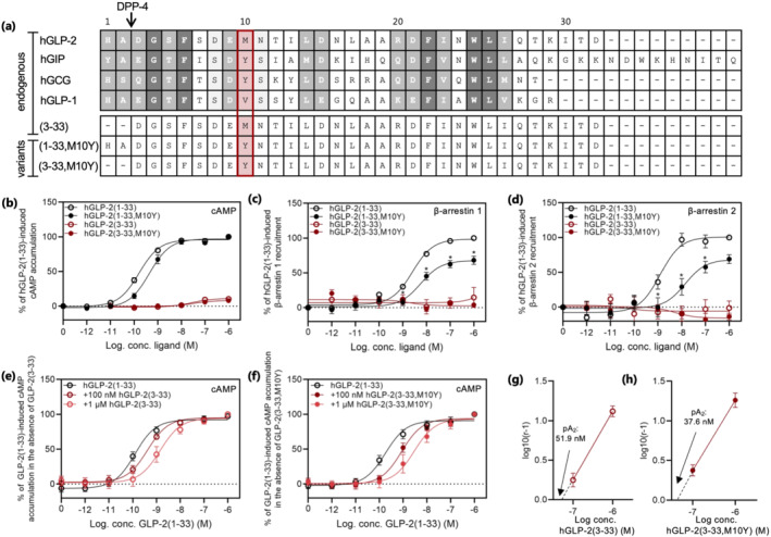
FIGURE 1.

Sequence alignment of GLP‐2 and related peptides and activity of hGLP‐2 and variants at the hGLP‐2 receptor. (a) Alignment of the class B1 GPCR peptides; hGLP‐2(1–33), hGIP(1–42), hglucagon (GCG)(1–29), hGLP‐1(7–36) (top panel) and the GLP‐2 variants; hGLP‐2(3–33), hGLP‐2(1–33,M10Y) and hGLP‐2(3–33,M10Y) (bottom panel). Dark grey refers to positions, which are fully conserved (identical); medium grey refers to positions with strongly similar residues, while light grey refers to positions with weakly similar residues. The red box marks position 10 (counted from residue 1 of hGLP‐2(1–33)). Dose–response curve in (b) cAMP accumulation, (c) β‐arrestin 1 recruitment and (d) β‐arrestin 2 recruitment for the hGLP‐2 receptor stimulated with increasing concentrations of hGLP‐2(1–33) and, hGLP‐2(1–33,M10Y), both in black, and, hGLP‐2(3–33), and hGLP‐2(3–33,M10Y), both in red. cAMP accumulation dose–response curve for hGLP‐2(1–33) in the presence of 100 nM and 1 μM (e) hGLP‐2(3–33) or (f) hGLP‐2(3–33,M10Y), and the corresponding Schild plots shown in (g) hGLP‐2(3–33) and (h) hGLP‐2(3–33,M10Y) with pA2‐values. To compensate for inter‐assay variations data were normalized to 1 µM hGLP‐2(1–33)‐mediated activation and recruitment within each assay. Potencies, efficacies and number of experiments are included in Table 1
