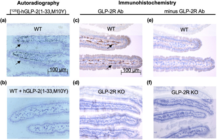
FIGURE 6.

Autoradiography and immunohistochemistry in mice. Histological sections of the small intestine after (a, b) autoradiography in mice injected with [125I]‐hGLP‐2(1–33,M10Y) for (a) wild type (WT) mice and (b) WT mice pre‐injected with >10,000‐fold excess unlabelled hGLP‐2(1–33,M10Y) and (c, f) immunohistochemistry using a polyclonal rabbit GLP‐2 receptor (R) antibody in (c) WT mice and (d) GLP‐2 receptor KO mice. In the absence of the primary GLP‐2 receptor antibody, the secondary biotinylated goat‐anti rabbit antibody revealed no staining of either (e) WT or (f) GLP‐2 receptor KO mice. Scale bar 100 μm
