
Figure 2.
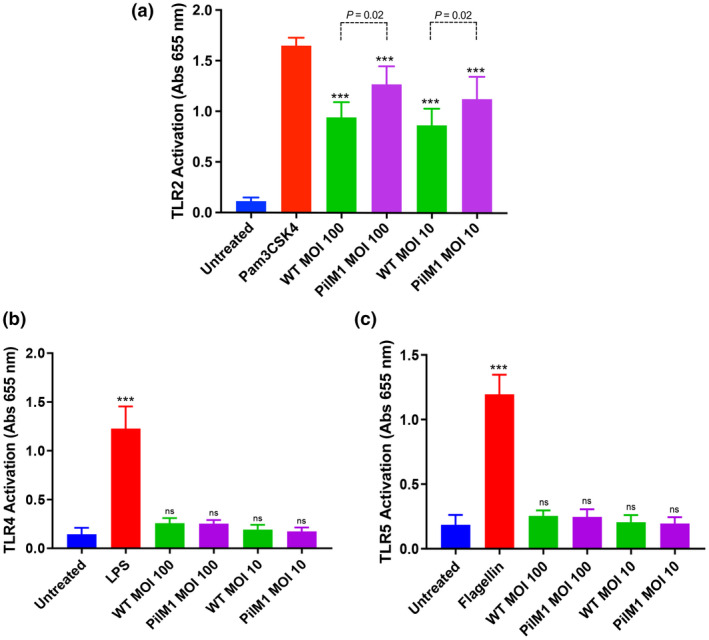
TLR2 is activated in response to the M1/T1 group A Streptococcus pilus. (a) HEK‐Blue hTLR2, (b) HEK‐Blue hTLR4 and (c) HEK‐Blue hTLR5 cells were incubated with wild‐type (WT) or PilM1 Lactococcus lactis strains, positive control (1 μg mL−1 Pam3CSK4 for TLR2, 100 ng mL−1 lipopolysaccharide for TLR4 or 1 μg mL−1 flagellin for TLR5) or left untreated. Levels of TLR activation were determined by absorbance at 655 nm. Comparisons with negative control denoted on experimental groups. Average of three independently performed experiments is shown. Experiments were performed in duplicates and data are shown as mean ± s.d. Statistical significance was determined by one‐way ANOVA, and P‐values were calculated by Holm–Šídák’s multiple comparisons test. ***P ≤ 0.001 compared with the negative control. MOI, multiplicity of infection; ns, not significant; s.d., standard deviation; TLR, Toll‐like receptor.