
Figure 1.
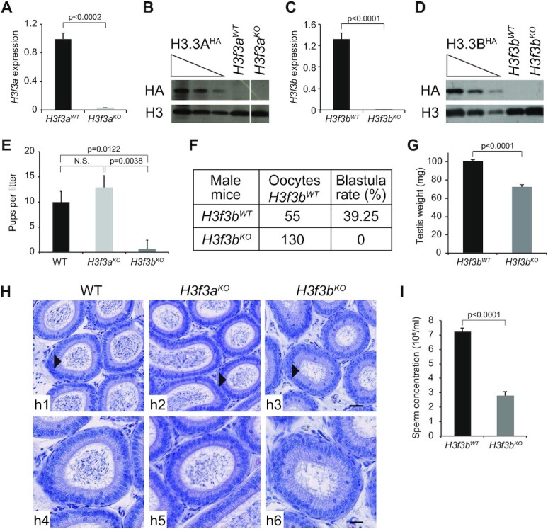
H3f3bKO, but not H3f3aKO mice, exhibit decreased sperm production and severe infertility. (A–D) Efficient depletion of H3.3A and H3.3B in the testis of the indicated knock-out mice. qPCR measurement of the mRNA level (A, C) and Western blotting (anti-HA) analyses (B, D) of either H3.3A (A, B) or H3.3B (C, D). (A, C) The average values of the mRNA of three biologically independent experiments are shown. (B, D) Total cellular lysates derivedfrom testis extracted from H3f3aHA, H3f3a WT and H3f3a KO mice (B) or H3f3bHA, H3f3b WT and H3f3b KO mice (D) were submitted to Western blot analysis. (E) Numbers of pups per female OF1 mated with either H3f3bWT, H3f3a null or H3f3b null male. The average of 9 litters is presented for 3 males by genotype. (F) Number of 2-cell and blastocysts obtained by in vitro fertilization using sperm from either control or H3f3b null males and oocytes prepared from control H3f3bWT females. (G) Testis weight for control and H3f3b null males. The average of four testes of different genotypes was presented. (H) Hematoxylin-stained cross-sections of epididymis of control (h1, h4), H3f3aKO (h2, h5) and H3f3bKO (h3, h6) male mice; h4–h6 show the enlarged epididymis cross-sections indicated by an arrow head in h1–h3; scale bars, 30 μm (h3) and 70 μm (h6). (I) Sperm concentration (millions per milliliter) for control H3f3bWT (n = 5) and H3f3b null males (n = 2). (A, C, E, G, I) P-values were calculated from Student's unpaired t-tests.