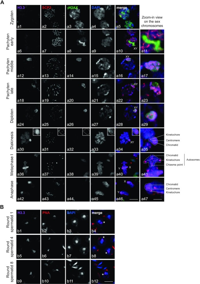
Figure 3.
H3.3B localized on the sex chromosomes from the middle pachytene to round spermatids during spermatogenesis. (A, B) Immunostaining of spermatogenic cells from meiosis (A) to post-meiosis (B). Cells were triple-stained on WT testis section with anti-H3.3, anti-γH2AX, anti-SCP3 (A) or anti-PNA (B) as well as DAPI. Developmental stages were classified by staining profiles of SCP3, γH2AX and PNA. Scale bars, 8 μm (a46), 3 μm (a47) and 8 μm (b12). The asterix represents the sex chromosome either x or y (b4, b8, b12).

