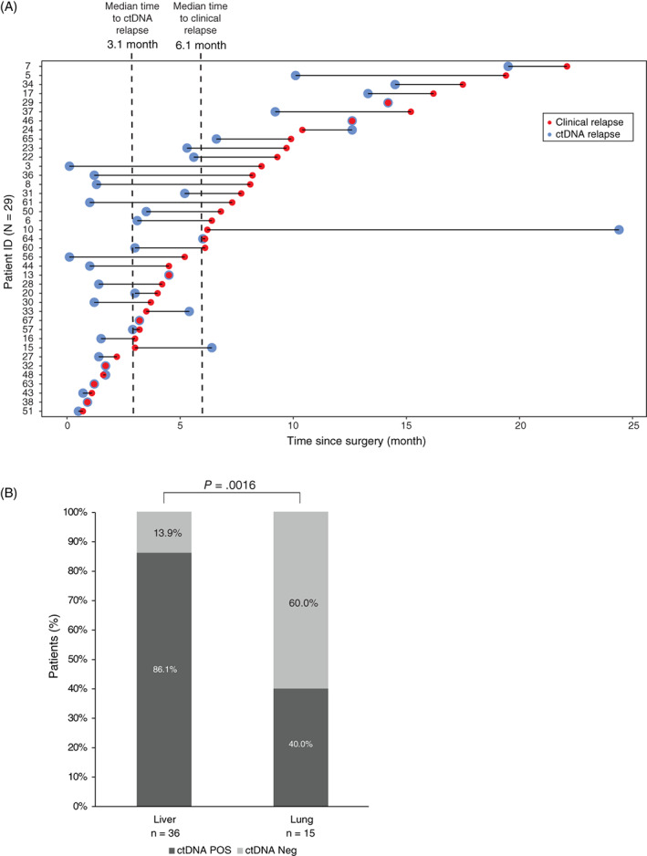
FIGURE 3.

Associations between ctDNA relapse and clinical relapse and between ctDNA status and recurrence location at time of relapse. (A) Patients are sorted by time to recurrence. Only patients with blood drawn prior to or at the day of CT imaging are included (N = 39). Patient ID 55 was excluded because the first longitudinal blood sample was drawn 2.8 months subsequent to the relapse. (B) ctDNA positivity of matched blood samples from patients with liver or lung metastases were unevenly distributed (Fisher's exact test, P = .0016)
