Figure 3.

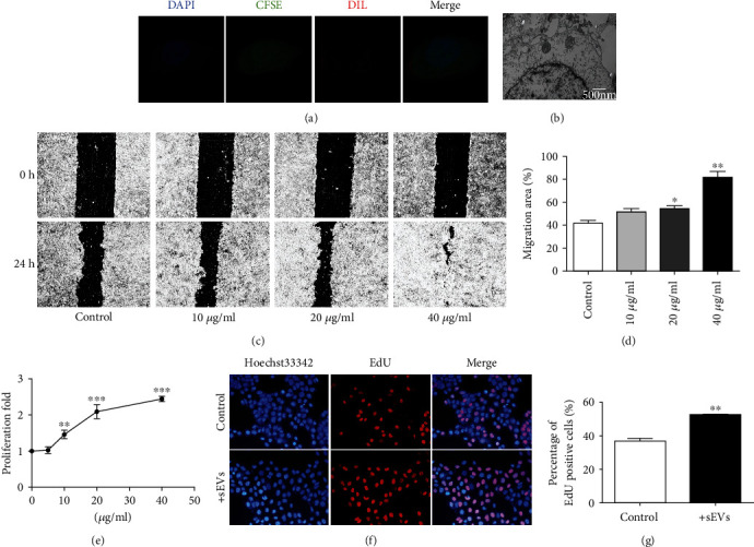
The effect of HUMSC-sEVs on HCECs' proliferation and migration in vitro. (a) Fluorescence images of CFSE-labeled HCECs (green) incubated with Dil-labeled HUMSC-sEVs (red). Nuclei were stained with DAPI (blue). (b) TEM of HCECs incubated with HUMSC-sEVs. (c, d) Representative images from in vitro scratch wound healing assays demonstrating that cell migrates into the cell-free region is significantly promoted in the presence of HUMSC-sEVs when compared to controls, n = 4. (e) CCK-8 assay showed increased proliferation of HCECs incubated with HUMSC-sEVs after 48 hours, n = 5. (f, g) The proliferating HCECs were detected by EdU incorporation. The cells were treated with HUMSC-sEVs or blank control, n = 3. Blue: nuclear staining (Hoechst33342); red: EdU staining. Data are expressed as the means ± SD. ∗P < 0.05, ∗∗P < 0.01, and ∗∗∗P < 0.001.
