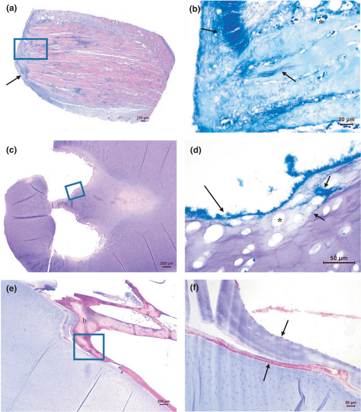
FIGURE 6.

Histopathology of ulcers on the head. (a, b) Skin and muscle tissues with filamentous rods, especially numerous towards not only on the surface but also present in endomysium between muscle cells, as well as within myocytes (arrows). Lack of inflammatory cells is evident. Dilated vessels as a sign of hyperaemia are seen (asterisk). (a: H&E, b: May‐Grünwald‐Giemsa (MGG) of a new section covering approximately the boxed area in a). (c, d) Mandibular cartilage with long filamentous rods present on irregular eroded surfaces of luminal structures (long arrow). Bacteria are also present within the cartilage (short arrows). The cartilage matrix is degenerated with enlarged lacunae devoid of chondrocytes (asterisk). (c: H&E, d: MGG of an area approximately covering the boxed area in c). (e, f) Numerous filamentous rods present in connective tissue in the interface between the mandibular cartilage and adjacent bone. Bacteria are also seen on the surface of the bone (b), probably associated with the fibrous layer covering the bone (arrows). (e: H&E, f: IHC of an area approximately covering the boxed area in e)
