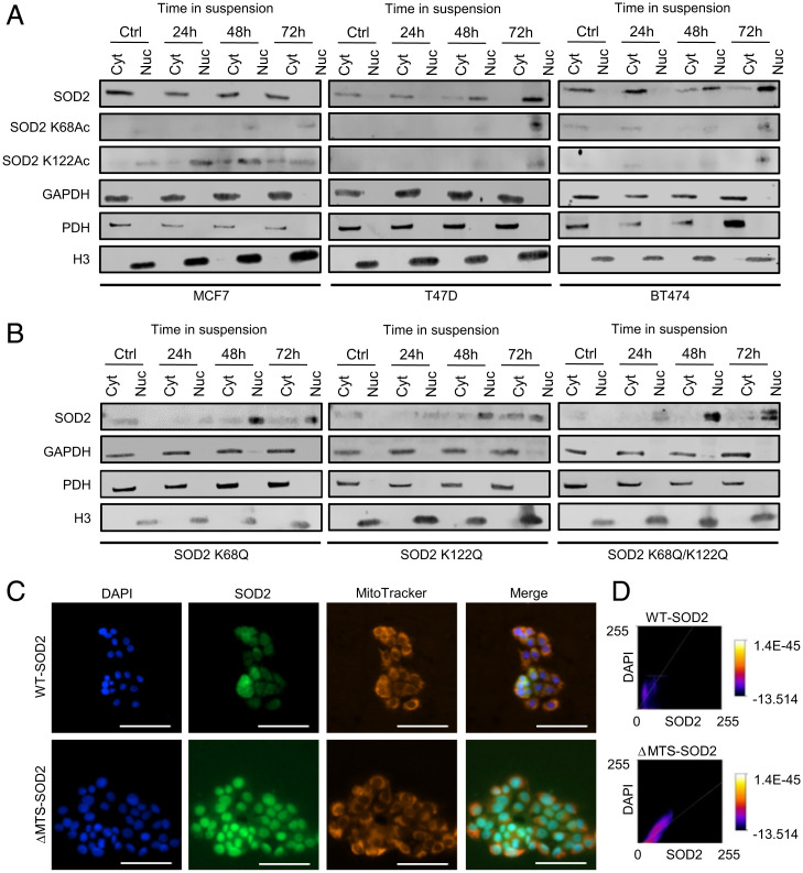
Fig. 1.
Acetylated SOD2 localizes to the nucleus. (A) Acetylated SOD2 accumulates in the nuclear fraction of breast cancer cell lines cultured in suspension. (B) SOD2 accumulates in the nuclear fraction of MCF7 cells expressing SOD2 K68Q, K122Q, and K68Q/K122Q mutants. (C) SOD2 localizes to the nucleus in the absence of MTS. (D) Quantification of WT- and ΔMTS-SOD2 colocalization with nuclear staining. Figures are representative of at least three independent experiments. Cyt, extranuclear fraction; Nuc, nuclear fraction; GAPDH, glyceraldehyde 3-phosphate dehydrogenase–cytosolic subcellular localization marker; PDH, pyruvate dehydrogenase–mitochondrial subcellular localization marker; H3, histone 3–nuclear subcellular localization marker. (White scale bars, 100 µm.)

