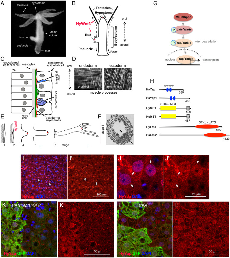
Fig. 1.
Hydra homolog of YAP is expressed in ectodermal epithelial cells. (A) Photo of a live Hydra. (B) Schematic of Hydra body plan; arrows indicate directions of cell displacement along the oral/aboral axis. (C) Schematic of a section through the Hydra body column. (D) Hydra endodermal and ectodermal muscle processes visualized with phalloidin. (E) Schematic of Hydra budding (adapted from ref. 11). The area of expression of HyWnt3 is confined to about 50 ectodermal epithelial cells marked in red (12). (F) Stage 1: Transverse section through the budding zone, arrows indicate increased cell density in the ectoderm and endoderm (adapted from ref. 13). (G) Schematic of the Hippo pathway. (H) Schematic of Hydra and mammalian homologs of Yap, MST, and LATS proteins; WW - proline-rich sequences binding domain; STKc–MST1/2 - catalytic domain of MST family of serine/threonine kinases;  - MST1-SARAH–apoptosis-mediating domain; STKc–LATS - catalytic domain of LATS family of serine/threonine kinases. (I–J′) Apical view of Hydra ectoderm immunostained with anti-HyYap serum. Arrows point to the nuclei of ectodermal epithelial cells. (K–L′) Apical view of ectoderm of GFP polyp electroporated with shGFP/HyYap (K and K′), shGFP alone (L and L′) hairpins and immunostained with anti-GFP and anti-HyYap antibodies; animals were fixed 6 d after electroporation.

