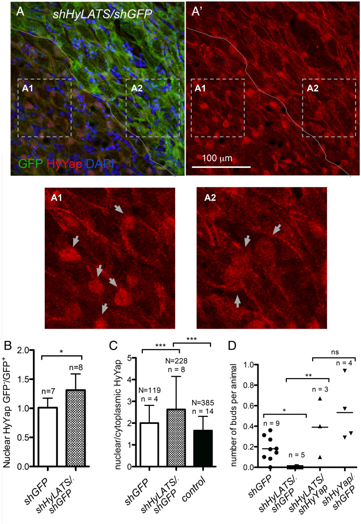
Fig. 3.
HyLATS regulates cellular localization of HyYap and the budding rate. (A, A′, A1, and A2) Apical view of Hydra ectoderm electroporated with shHyLATS/shGFP and immunostained for GFP and HyYap; the border between GFP+ and GFP− areas is marked; arrows point to nuclei; A1 and A2 are high magnification of GFP− and GFP+ areas. Nuclear abundance of HyYap increases in GFP− ectodermal epithelial cells. (B) Graph shows the ratio of nuclear HyYap intensities between GFP− and GFP+ areas of GFP polyps electroporated with either shGFP or shGFP/shHyYap. Note, that electroporation with shGFP alone does not affect the nuclear abundance of HyYap (GFP−/GFP+ ∼1). The average intensities of immunostaining were measured and the ratios were calculated individually for each animal; n, number of polyps; two-tailed unpaired t test. (C) Graph shows the ratio between nuclear and cytoplasmic HyYap in nonelectroporated GFP+ ectodermal epithelial cells, cells electroporated with shGFP alone, and cells electroporated with shGFP/shHyLats. Nuclear/cytoplasmic ratio was measured and calculated for each individual cell. N, number of cells; n, number of polyps; two-tailed unpaired t test. (D) Graph shows the budding rate of hydras electroporated with either shGFP (control), shHyLats/shGFP, shHyLats/shHyYap, or shHyYap/shGFP. n, number of experiments; 10 to 20 polyps were used in each experiment for each condition; two-tailed unpaired t test. P values are: ns (P > 0.05), *(P ≤ 0.05), **(P ≤ 0.01), ***(P ≤ 0.001).

