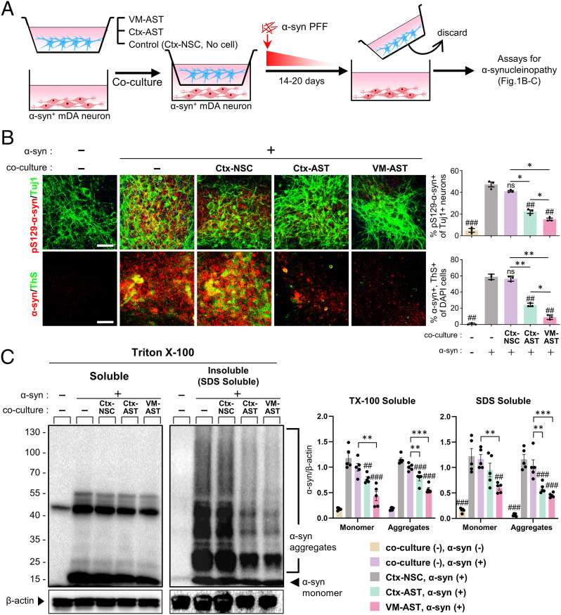
Fig. 1.
Cultured astrocytes reduce the aggregated and monomer forms of α-syn protein in the in vitro α-synucleinopathic PD cellular model. (A) Schematics of the coculture experimental procedures. Astrocytes derived from the VM or Ctx of rat pups (undifferentiated Ctx-NSCs as a control; SI Appendix, Fig. S1 A–C) were cocultured with α-syn–overexpressing mDA neurons (SI Appendix, Fig. S1 D and E) using the culture insert (1 x10⁵ cells plated each in the upper and lower chambers, separated using a polycarbonate membrane with 0.4-µm pore size). As another control, the culture insert without cell seeding was used. Media containing sonicated α-syn PFF (1 µg/mL) was added to both the upper and lower compartments and followed by gradual dilution of α-syn PFF by changing half the medium every other day for 14 to 20 d. (B) α-syn aggregation detected by immunoreactivity against pS129-αsyn and Thioflavin S staining. (C) WB-based detection of monomer and aggregate forms of α-syn in the Triton X-100–soluble and –insoluble (SDS soluble) fractions. Significant differences from the α-syn-treated (+) and without cocultured cells (−) control at ##P < 0.01 and ###P < 0.001 and between the groups indicated at *P < 0.05, **P < 0.01, and ***P < 0.001. n = 3 (B) and 5 (C), one-way ANOVA, followed by Bonferroni post hoc analysis. ns, no significance. (Scale bars: 25 μm.)

