
Fig. 1.
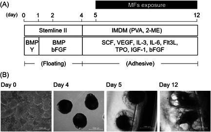
Differentiation of hiPS cells into HSPCs, and MF exposure during differentiation from mesodermal cells to HSPCs. (A) Schematic of the protocol for hematopoietic differentiation and MF exposure. (B) Morphology of the cells at each period. After EBs (day 4) were formed from hiPS colonies (day 0), the EBs were attached onto gelatin‐coated culture dishes (day 5). Exposure of MFs was performed for 7 days until day 12. Scale bars represent 1000 μm. EB = embryoid body; hiPS = human‐induced pluripotent stem; HSPC = hematopoietic stem progenitor cells; MF = magnetic field.