FIGURE 1.

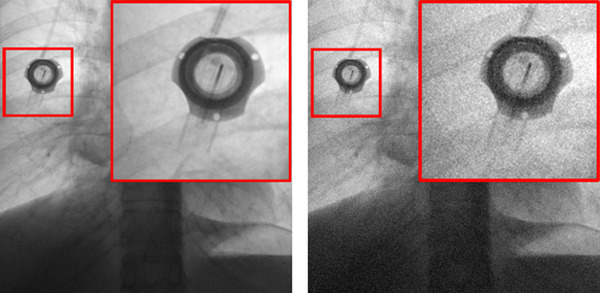
An example of adding simulated noise to a chest X‐ray image. Left: Image acquired at a relatively high dose. Right: Image with 60% simulated added noise

An example of adding simulated noise to a chest X‐ray image. Left: Image acquired at a relatively high dose. Right: Image with 60% simulated added noise