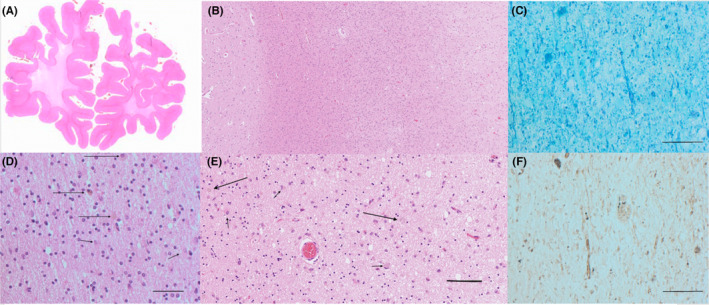
FIGURE 2.

Histopathology of patient 2 postmortem brain tissue sections. (A) Macrophotograph anterior frontal lobes, hematoxylin–eosin staining. Note the well‐delineated cortical ribbon, which is of normal width and the subjacent white matter, which is severely attenuated, with shrinkage of the entire frontal lobes (B) Microphotograph posterior frontal lobe, hematoxylin–eosin. Note the intact cortical tissue with dense and preserved matrix (left) and the relatively preserved subcortical white matter (center), while deeper white matter is markedly attenuated (right) (C) Microphotograph anterior frontal lobe, Luxol Fast Blue‐Cresyl violet. Note the extreme scarcity of myelin sheaths and the axonal and myelin sheath swelling in one of the few remaining long structures. D: White matter of the posterior frontal lobes, hematoxylin–eosin. Note the attenuated tissue with many reactive astrocytes and a few mildly pigmented macrophages. (E) Pleomorphic reactive astrocytes and macrophages. (F) Attenuated white matter from the anterior frontal lobe, as in A and C, Gallyas silver staining for identification of axons. Note the rudimental fragments of axons and the two macrophages (pigmented) with granular intracellular debris. Bar indicates 0.05 mm
