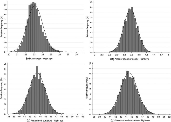
FIGURE 3.

Distribution of ocular biometry parameters of the right eye in a sub‐group of the sample (n = 7901; consisting of all children in the first two schools, and all children with a refractive error requiring correction plus all emmetropic children from grades 1, 4 and 6 from the other nine schools) for (a) Axial length (in mm); (b) Anterior chamber depth (in mm); (c) flattest corneal meridian (K1) (in dioptres) and (d) steepest corneal meridian (K2) (in dioptres)
