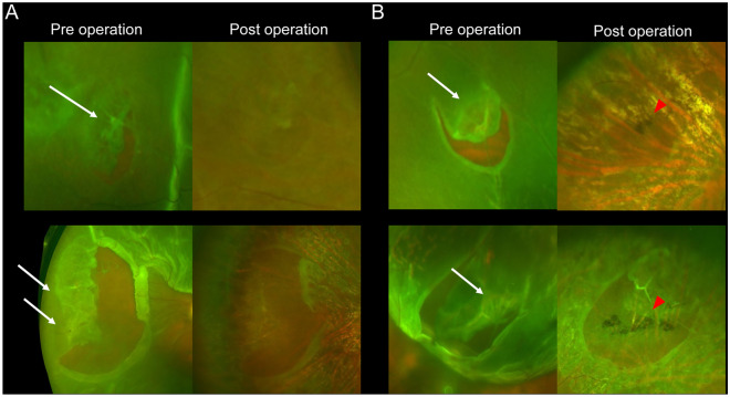
Figure 1.
Lattice degeneration (LD) associated with rhegmatogenous retinal detachment (RRD) examined by pre- and postoperative ultra-widefield (UWF) color scanning light ophthalmoscopy (SLO). (A) LD without a pigmentary lesion (PL) in two representative cases. (B) LD with PL in two representative cases. The preoperative UWF images show the white vessels and retinal thinning within the LD lesions. White arrows indicate LD, and red arrowheads indicate PL.

