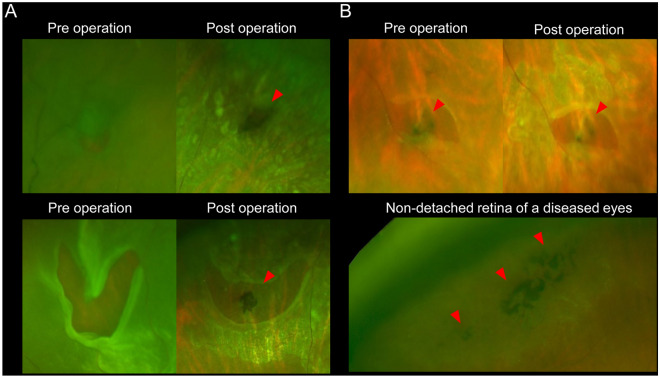
Figure 2.
Pigmentary lesion (PL) of eyes with rhegmatogenous retinal detachment (RRD) examined by pre- and postoperative ultra-wide field (UWF) scanning light ophthalmoscopy (SLO). (A) Pigmentary lesion (PL) without lattice degeneration (LD) in representative two RRD-eyes. The preoperative UWF-SLO images do not clearly show the PL. However, the postoperative UWF-SLO images show the PL in and behind the retinal tears, at the retinal pigment epithelial level. In the eyes, the retinal tears do not accompany the LD lesion. (B) PL without LD in other RRD-eyes. The pre- and postoperative SLO images show the PL. Red arrowheads indicate PL.

