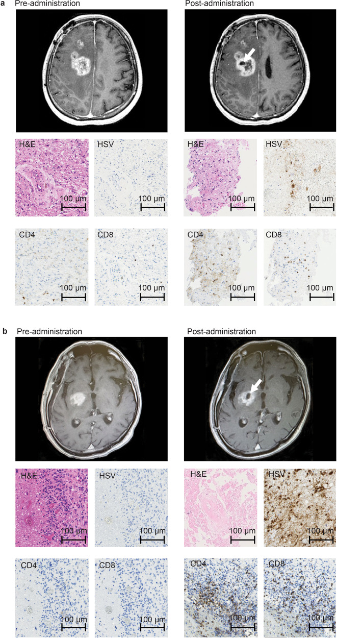
Fig. 2. MRI and histology studies in two patients (a: Patient #6 and b: Patient #10).
MRI showed enlargement of the entire enhanced lesion with clearing of contrast enhancement at the injection site (arrows) post-G47Δ administration. All specimens from pre-administration were negative for HSV-1 immunostaining. Second biopsy specimens obtained before the second injection from the same coordinates of the first G47∆ injection 7 days (a) and 6 days (b) post-administration showed decreased number of tumor cells, possibly due to tumor cell destruction associated with viral replication (H&E) and positive HSV-1 immunostaining. An increase in infiltration of CD4+ and CD8+ T lymphocytes towards remaining tumor cells was observed. Pathology images are representative of four biopsy specimens.

