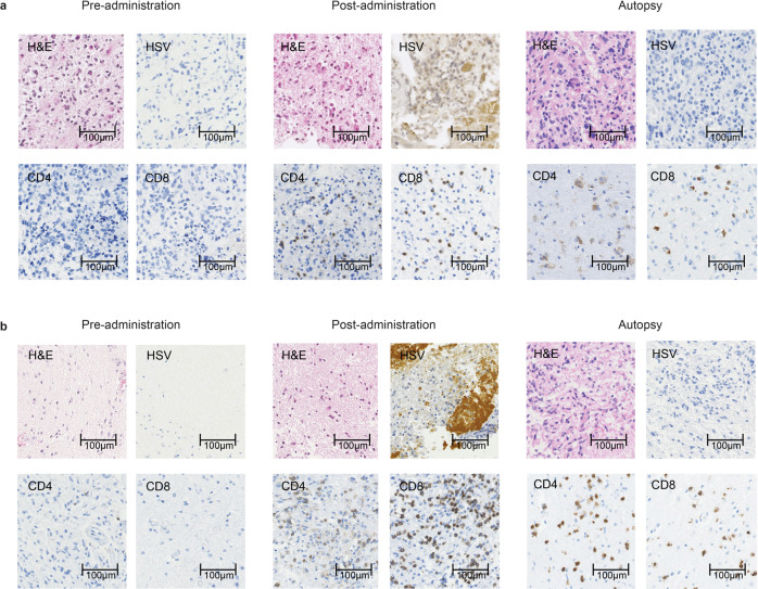
Fig. 5. Histological findings in autopsy cases.
a Patient #11: 6.2 months after receiving G47Δ and b Patient #13: 13.7 months after receiving G47Δ. Both brain tumor specimens from pre-administration showed negative immunostaining for HSV-1, and very scarce CD4+ and CD8+ T lymphocytes. Infiltration of CD4+ and CD8+ T lymphocytes and positive HSV-1 immunostaining were observed after G47∆ administration at the injection site in both cases. Representative of four biopsy specimens. At autopsy, both brain tumor specimens showed viable glioblastoma cells (H&E) together with intratumoral infiltration of CD4+ and CD8+ T lymphocytes. The infiltration of CD4+ and CD8+ T lymphocytes was shown to persist for >13 months. Autopsy brain tumor specimens were negative for HSV-1 immunostaining. Representative of three tissue samples.

