
Fig. 16.
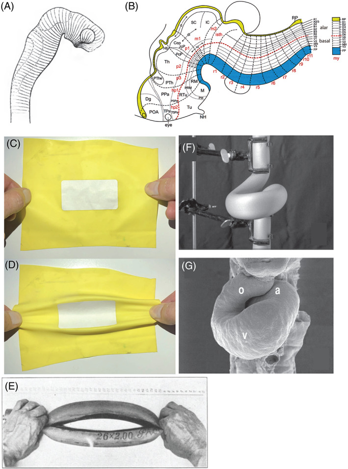
(A) His's model of longitudinal zones in the neural tube. Fig. 13 from His (1888b). (B) The ‘updated prosomeric’ model of brain organisation. Yellow, roof plate; blue, floor plate. Fig. 8 from Puelles (2018). (C, D) Folding along elasticity boundaries as a mechanism of early vertebrate morphogenesis (reconstructed by Dr. Fleury, based on fig. 16 in Fleury et al., 2015). A paper sticker is placed on a rubber sheet, thereby stiffening the central area. The stretched sheet buckles around the sticker, along boundaries between elastic and stiff regions. This demonstration is reminiscent of His' suggestion that the early embryo has variable elasticity and undergoes folding at unstable boundaries. (E) Vaihinger modelled the opening and closing of plant stomata by pushing and pulling a rubber tube (fig. 8 from Vaihinger, 1941). Courtesy of Universiteitsbibliotheek Utrecht. (F, G) Manipulation of a rubber tube (F) can create shapes resembling a scanning electron micrograph (G) of the developing cardiac tube of the chicken (fig. 5B, D from Männer, 2004).