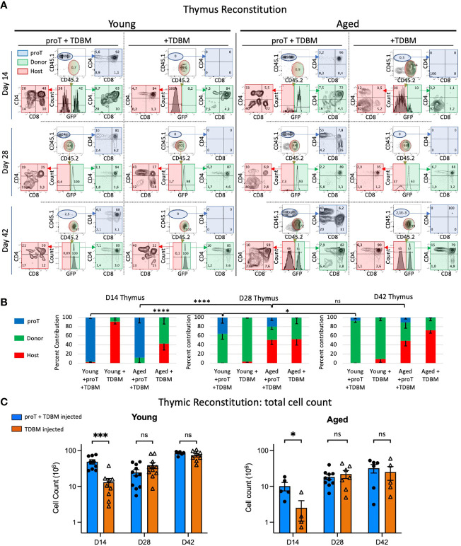
Figure 4.
Thymic engraftment of young and aged mice. Young (8-12wks) and aged (18-20 months) mice were treated with CTX followed by lethal irradiation in preparation for i.v. injection of either 4 x 10⁶ proT cells derived from LSK/OP9-DL4 cocultures plus 1 x10⁶ TDBM or 1 x10⁶ TDBM alone as indicated. Thymus and other organs were harvested on days 14 (D14), D28 and D42. Thymocytes were labelled with appropriate lineage markers and analyzed by flow cytometry represented by plots shown in (A). The thymocyte population was contributed by cells derived from the host (red), GFP+ BM graft (green) and proT coculture (blue). The percent contribution of the source, whether host (CD45.2+, GFP-), BM graft-derived (CD45.2+, GFP+) or proT-derived (CD45.1+) is shown in (B) for the three timepoints. Total cell number of thymocytes is depicted in (C) with young in the left panel and the aged in the right panel. For all samples at all time points, n≥4 up to 12. Error bars indicated SEM as indicated in Methods; significance was measured with two-way ANOVA. *p<0.05, ***p<0.001, ****p<0.0001.

