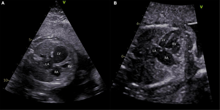
FIGURE 1.
Different phenotypes of left ventricular hypoplasia in the fetus. (A) A four-chamber view of a fetus with 24 weeks and critical aortic stenosis is shown with a severely dilated left ventricle. Panel (B) shows a four-chamber view of a fetus at 27 weeks with a long and skinny, but apex forming left ventricle. LA, left atrium; LV, left ventricle; RA, right atrium; RV, right ventricle.

