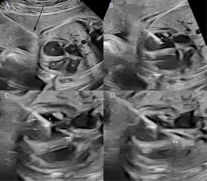
FIGURE 4.
Technique of fetal aortic valvuloplasty performed under ultrasound guidance. Fetal aortic valvuloplasty of a fetus at 25 weeks of gestation with critical aortic stenosis and eHLHS is shown. In panel (A) the ultrasound guided puncture of the fetal thoracic wall is shown, the arrow marks the needle just before entering the fetus. The needle has been introduced into the left ventricle (B), asterisk marks the needle tip inside the left ventricle. The guidewire and the balloon catheter have crossed the aortic valve in panel (C) and the needle is slightly retracted to allow good positioning of the balloon just before dilation of the aortic valve (**). Panel (D) shows full insufflation of the balloon catheter (**). eHLHS, evolving hypoplastic left heart syndrome; LA, left atrium; LV, left ventricle; RA, right atrium; RV, right ventricle.

