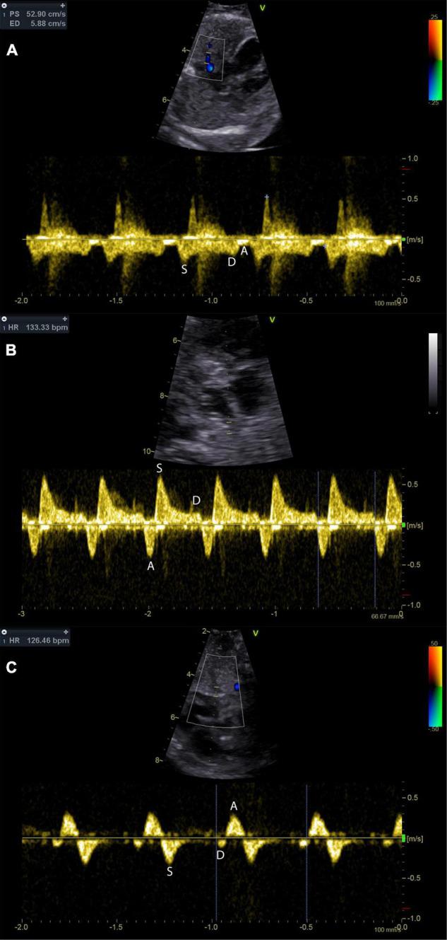
FIGURE 5.

Pulmonary vein Doppler patterns in the hypoplastic left heart with incremental restriction of the interatrial communication. Panel (A) shows a fetus with CAS and an unrestrictive interatrial communication; note that there is reduced flow during atrial systole (A-wave). Panel (B) shows a fetus with HLHS and restrictive, but patent atrial septum. Prominent A-wave flow reversal during atrial systole may be indicative of relevant restriction. Panel (C) shows the pulmonary venous flow pattern in a fetus with HLHS and intact atrial septum. Diastolic pulmonary vein flow is almost missing.
