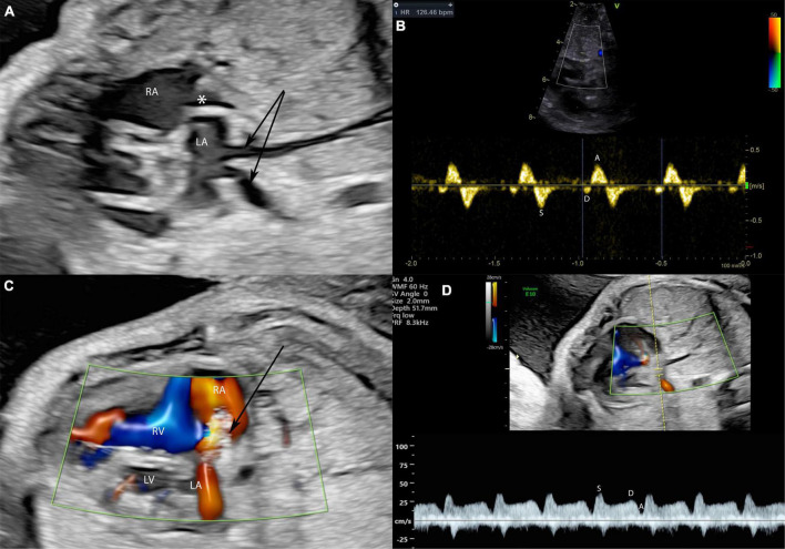
FIGURE 6.
A fetus at 28 weeks gestation with HLHS and intact atrial septum undergoing FCI. Stenting of the interatrial septum in a fetus with intact atrial septum and HLHS is shown. In panel (A) severely dilated pulmonary veins (arrows) indicate severe left atrial hypertension. The asterisk marks a thickened and into the right atrium bowing atrial septum. In panel (B) severe left atrial hypertension is indicated by severely abnormal pulmonary vein Doppler with almost missing diastolic flow and severe A-wave reversal during atrial systole. The arrow in panel (C) marks the placed atrial septum stent with good flow through the stent indicated by the red color Doppler signal. Note that the pulmonary vein Doppler in panel (D) is almost normalized in the same patient directly after stent placement with good diastolic flow and no presence of A-Wave reversal.

