
FIGURE 2.
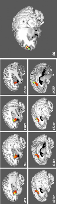
Comparison overlays between the cortical parcellation data (green) and ALE data (red) from Figure 1 in the left cerebral hemisphere. Regions were visually assessed for inclusion in the network if they overlapped with the ALE data. Parcellations included in the model of salience were identified in the insula, including AVI, FOP4, FOP5, and MI (top row); the middle cingulate gyrus, including a24pr, a32pr, p32pr, and SCEF (bottom row); and the dorsolateral prefrontal cortex, including 46 (middle). The labels indicate the parcellation shown in each panel. Abbreviations: a24pr, anterior 24 prime; a32pr, anterior 32 prime; AVI, areas anterior ventral insula; FOP4, frontal operculum 4; FOP5, frontal operculum 5; MI, middle insula; p32pr, posterior 32 prime; SCEF, supplementary and cingulate eye field