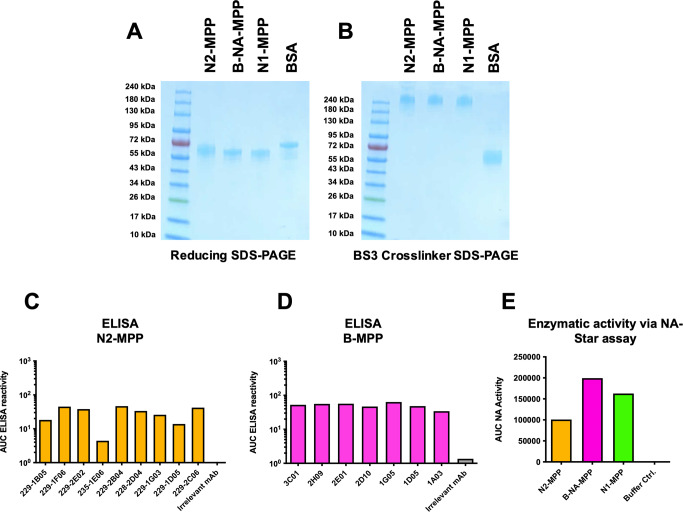
Fig. 6. Structural analysis of N2-MPP and B-NA-MPP.
A SDS-PAGE under denaturating conditions, all proteins show monomeric structures at an expected size of ~60 kDa. BSA was included as a monomer control. B SDS-PAGE using a BS3 crosslinker. N1-MPP, N2-MPP and B-NA-MPP show tetrameric structures at around 240 kDa. BSA was included as a monomer control. C ELISA against rec. N2-MPP using a broad panel of human anti-N2 mAbs to verify correct presentation of epitopes. D ELISA against rec. B-NA-MPP using a broad panel of human anti-B-NA mAbs to verify correct presentation of epitopes. E Enzymatic activity of N1-MPP, N2-MPP, B-NA-MPP was determined via NA-Star assay. Assays in C to E were run once in duplicates and the duplicates were used to calculate one area under the curve (AUC) value.

