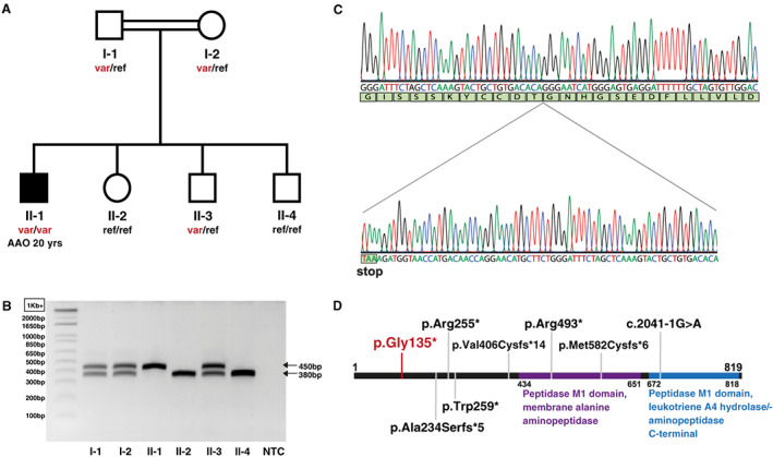
FIG 1.

Pedigree and genetic results. (A) The AOPEP variant is present in homozygous state in the subject affected by dystonia (black symbol) but in none of his unaffected relatives (white symbols). (B) Agarose gel electrophoresis of PCR fragments containing the AOPEP exon 2; wild‐type allele: 380 bp; variant allele: 450 bp. (C) Electropherogram showing the 70‐nucleotide duplication in AOPEP exon 2 in the DNA amplified from the affected subject. (D) Schematic representation of the AOPEP protein with known functional domains 4 and variants reported in patients with dystonia in recent studies (gray anchors) and in this study (in red). AAO, age at onset; NTC, negative control; ref, reference (wild‐type) allele; var, AOPEP c.333_402dup (p.Gly135*) variant allele. [Color figure can be viewed at wileyonlinelibrary.com]
