
FIGURE 2.
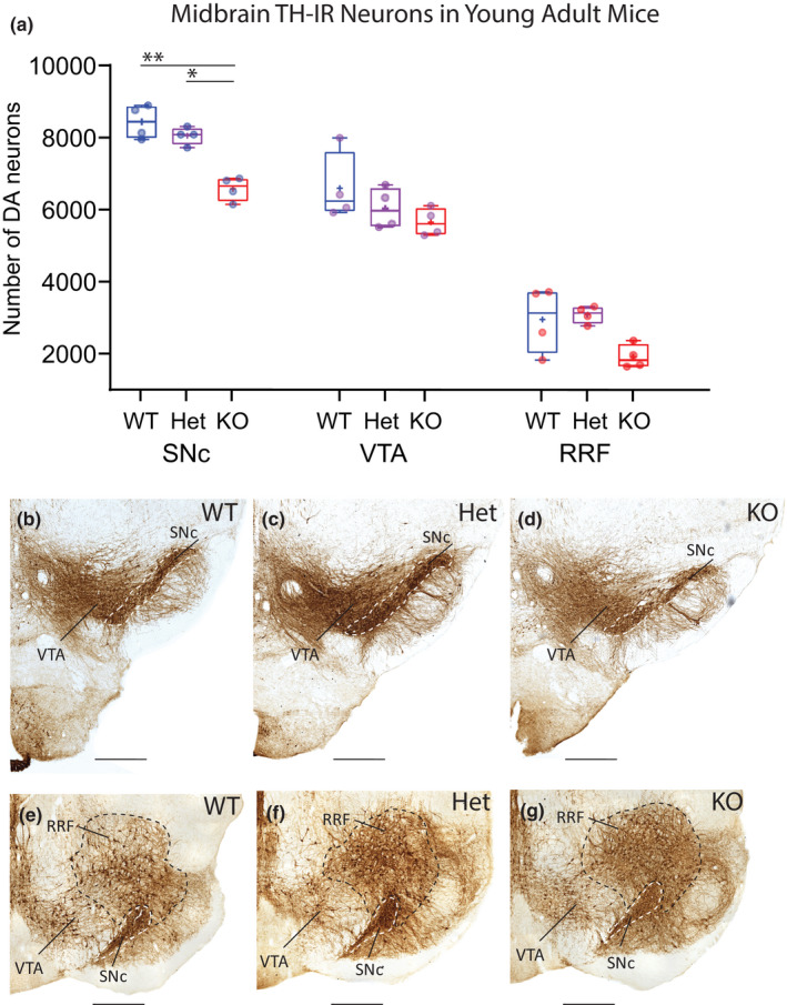
Reduced numbers of mDA neurons in SNc of young adult cDCC KO mice but not in cDCC Hets. (a) Comparison of stereological counts of mDA neuron subpopulations in 2.5‐month‐old young adult cDCC WT (DATCre/DCCwt/wt, n = 4), cDCC Het (DATCre/DCCfl/wt, n = 4) and cDCC KO (DATCre/DCC fl/fl, n = 4) mice. cDCC KO mice have significantly fewer mDA neurons in the SNc compared to either cDCC WTs (**p= 0.0018) or cDCC Hets (*p = 0.0207). The VTA and RRF contain similar numbers of mDA neurons in all 3 genotypes indicating that in young adults these neurons are spared from the impact of DCC loss. Statistical analysis applied is a two‐way ANOVA with Tukey’s HSD posthoc tests. Statistical significance was set as *p < 0.05, **p < 0.01, ***p < 0.001). Graphs are box and whiskers plots including all data points, median, mean (+ sign), 1st and 3rd quartile delimiting box, and minimum and maximum values at whiskers. n = number of animals. (b–g) The distribution and intensity of TH‐IR in the SNc (dashed boundary), VTA and RRF in all 3 genotypes is similar, and differences in mDA counts are discerned by quantitative methods using unbiased stereology. SNc, Substantia nigra pars compacta; VTA, ventral tegmental area; RRF, retrorubral field. Scale bars B‐G, 500 μm