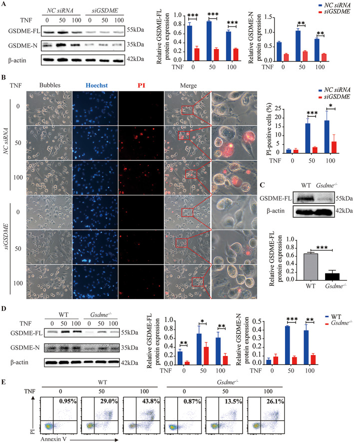
Figure 5.

Inhibition of tumor necrosis factor (TNF)–induced pyroptosis in macrophages by GSDME silencing. A and B, After transfection with GSDME or negative control (NC) small interfering RNAs (siRNAs) for 48 hours, THP‐1 cell–derived macrophages were left untreated or treated with 50 or 100 ng/ml TNF for the indicated times. A, Western blot of GSDME‐FL and GSDME‐N expression (left), quantification of GSDME‐FL expression (middle), and quantification of GSDME‐N expression (right) in macrophages treated as indicated for 36 hours after the silencing of GSDME. Values are the protein level relative to β‐actin. B, Left, Representative brightfield and fluorescence microscopy images showing the morphology of macrophages treated as indicated after the silencing of GSDME. For the merged images, right panels show higher‐magnification views (original magnification × 1600) of the boxed areas in the left panels (original magnification × 400). Right, Percentage of propidium iodide (PI)–positive cells. C, Western blot (top) and quantification (bottom) of GSDME‐FL expression in wild‐type (WT) and Gsdme −/− mice, showing the efficiency of Gsdme knockout. Values are the protein level relative to β‐actin. D, Western blot of GSDME‐FL and GSDME‐N expression (left), quantification of GSDME‐FL expression (middle), and quantification of GSDME‐N expression (right) in lysates of bone marrow–derived macrophages (BMMs) from WT and Gsdme −/− mice, cultured with the indicated concentrations of TNF for 36 hours. Values are the protein level relative to β‐actin. E, Flow cytometric analysis of murine BMMs treated as indicated and stained with annexin V/PI. In A, B, and D, bars show the mean ± SD. * = P < 0.05; ** = P < 0.01; *** = P < 0.001. See Figure 1 for other definitions. Color figure can be viewed in the online issue, which is available at http://onlinelibrary.wiley.com/doi/10.1002/art.41963/abstract.
