
FIGURE 7.
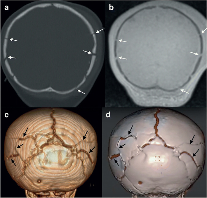
A 9‐month‐old with multiple skull fractures (arrows) demonstrated on coronal head computed tomography (CT) (a), and coronal black bone magnetic resonance imaging (MRI) (b) and the corresponding 3D rendering (c, d). Reprinted by permission from Springer Nature from reference 70.