
FIGURE 9.
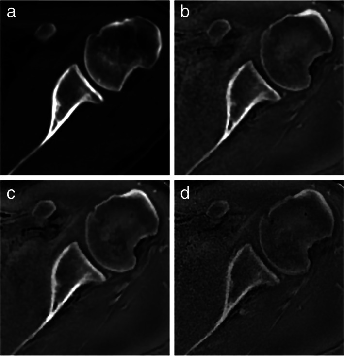
Axial computed tomography and zero echo time magnetic resonance imaging of left shoulder in a 38‐year‐old man. Axial images obtained by computed tomography (a) and zero echo time magnetic resonance imaging at 1.0 mm3 (b), 0.8 mm3 (c), and 0.7 mm3 (d) all show high‐contrast imaging of the osseous structures, including the glenoid and glenohumeral joint. Reprinted with permission from reference 19.