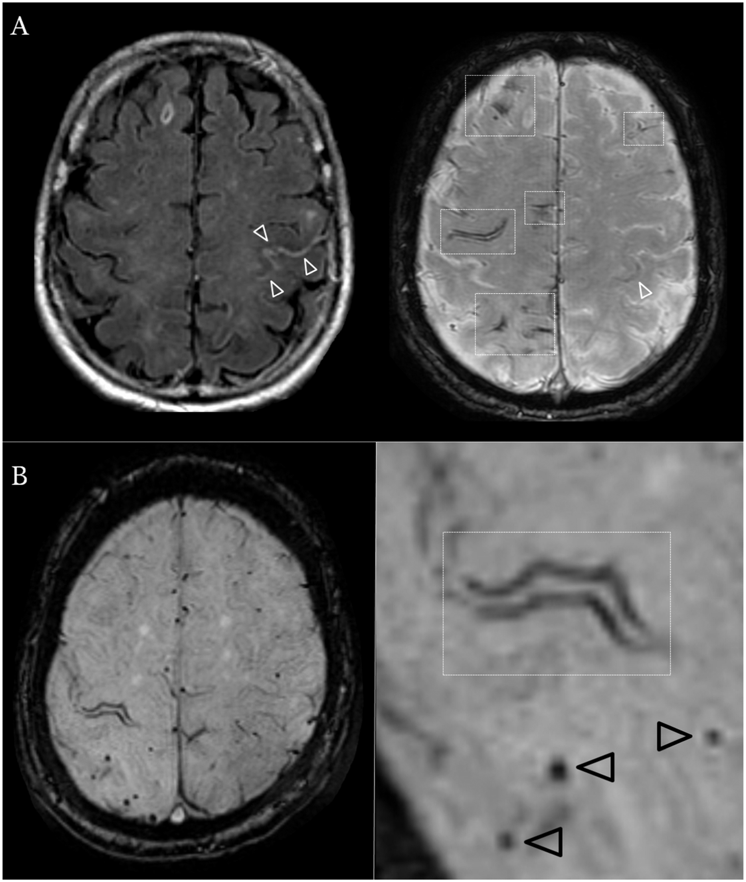
Figure 3.

Representative patients with and without cSAH.
1.5 Tesla axial brain MRI sequences (A) FLAIR (left) sequence and SWI (right) axial sections in an 82-year old male evaluated for rapidly progressing right sided tingling resolved in 15 minutes after onset. No relevant medical history. Imaging demonstrates acute subarachnoid blood in the central sulcus (white arrowheads) and bilateral disseminated foci of cortical superficial siderosis (white rectangles). (B) SWI axial sequence in a 75-year old female with repeated stereotyped episodes of dizziness and left sided hand tingling. Imaging demonstrates chronic focal cortical superficial siderosis affecting the right central sulcus (white rectangle) as well as strictly lobar cerebral microbleeds (black arrowheads).
