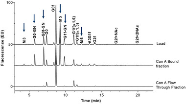
FIGURE 4.

Glycan peak identification of monoclonal antibody (mAb) by 2‐aminobenzamide (2‐AB)–HILIC. Glycan profiles among the load, concanavalin A (Con A)‐bound, and Con A flow‐through fractions were compared. Terminal‐mannose species present in original samples were not present in the flow‐through fractions, indicating that they were bound to the Con A column. Arrows indicate the presence of terminal mannose
